Imagine a surgical robot pausing — for a fraction of a second — to re-read a molecular signal from an engineered protein that has latched onto a tumor cell’s surface, then adjusting its instrument path by less than a millimeter to avoid clipping healthy nerve tissue. That scenario is not science fiction; it is the driving ambition behind a landmark collaboration now taking shape across the University of Texas System, where protein scientists, robotics engineers, and oncologists are working in deliberate concert for the first time.
A New Front in the War on Cancer Is Opening in Texas
Cancer remains the second leading cause of death in the United States, and the hard consensus among researchers is that incremental refinements to existing therapies are no longer sufficient on their own. The disease is too heterogeneous, its borders too unpredictable, and its molecular behavior too variable for any single discipline to solve it alone. That recognition is what makes the current moment in Texas significant: MD Anderson Cancer Center and UT Austin have launched a joint initiative to advance breakthroughs in cancer research, merging expertise in AI modeling, surgical robotics, and cancer therapeutics under a shared institutional framework. What follows explains what that convergence actually means mechanistically, why Texas became the hub for it, and what patients can — and cannot — realistically expect in the years ahead.
The Problem Precision Robotics Is Solving in Oncology
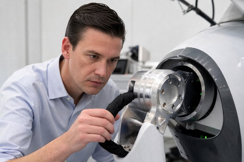
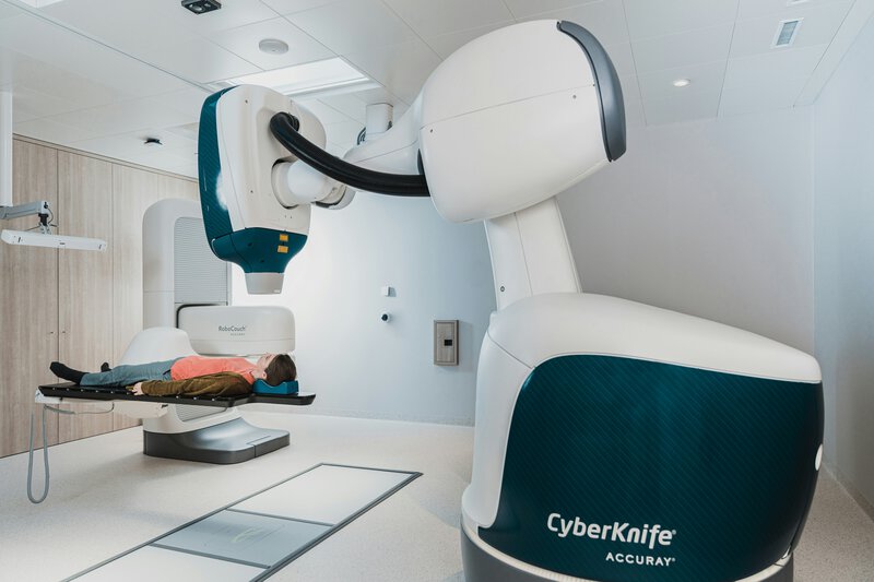
Precision robotics, as the term is used in surgical oncology, refers to robotic systems engineered to perform or assist in procedures with accuracy measured in fractions of a millimeter, materially reducing collateral damage to healthy tissue compared with conventional open or laparoscopic surgery. The da Vinci Surgical System and its successors are the most commercially familiar examples, but the broader category is evolving rapidly.
The core oncological challenge these systems confront is biological, not merely mechanical. Tumors are rarely uniform, cleanly bordered masses. They invade surrounding tissue unpredictably, their density varies across a single lesion, and their margins — the edges a surgeon must excise completely to minimize recurrence risk — are frequently invisible to the naked eye and inconsistently visible even under standard imaging. Getting those margins wrong carries direct clinical consequences: residual cancer cells left behind after surgery are a documented driver of local recurrence.
Evidence from the surgical literature, including studies published in The Lancet Oncology, has established that robotic-assisted surgery already reduces blood loss and shortens recovery time in several cancer procedure categories compared with conventional approaches. Whether those advantages translate into improved long-term survival outcomes remains an active and unresolved area of research — a distinction that honest science communication requires acknowledging plainly. What is not contested is the gap the Texas initiative is targeting: current robotic systems respond to what surgeons can see. The next generation, researchers argue, must respond to what surgeons cannot see — molecular-level data, updated in real time. Closing that gap requires protein science to enter the operating room.
Protein Engineering: The Molecular Intelligence Behind the Machines
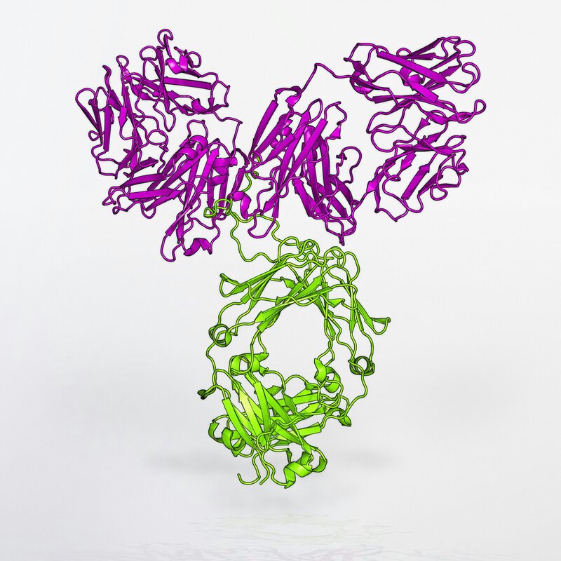
Protein engineering is the deliberate redesign or construction of proteins — the molecular machines responsible for virtually every biological function in living organisms — to perform tasks that nature never assigned them. In an oncology context, that might mean designing an antibody-like protein that binds exclusively to a surface marker found on a specific type of tumor cell while ignoring the chemically similar markers on adjacent healthy cells.
The mechanism linking protein science to surgical robotics is elegant in principle. Engineered proteins, including modified antibodies and purpose-built biosensors, can be designed to fluoresce — to emit detectable light — when and only when they encounter their target tumor tissue. A robotic imaging system equipped to detect that fluorescence would effectively have a molecular map of the cancer’s true boundaries, not just its visible contours. The surgeon, or an AI system guiding the robot, would be operating with information that currently does not exist in the operating room.
It is important to distinguish what is established from what remains emerging. Fluorescence-guided surgery using protein-based probes is a genuine and active clinical research area; trials are underway at several major cancer centers, and some fluorescence agents have received regulatory review. However, the integration of those probes with fully autonomous or AI-driven robotic platforms remains largely pre-clinical — a meaningful gap between promising components and a validated integrated system. Researchers within the UT System are partnering to span exactly this translational gap, from engineered protein probe to AI-interpreted surgical signal — a combination that, if validated in clinical trials, could redefine what “clean margins” means in oncological practice.
How Artificial Intelligence Ties Robotics and Biology Together
Artificial intelligence — specifically deep-learning models trained on large datasets of tumor imaging, molecular profiles, and surgical outcomes — functions in this system as an interpreter. The fluorescent signal from a protein probe is data; it is the AI layer that translates that data into actionable, real-time guidance for a robotic surgical instrument. Without that interpretive step, a surgeon still faces the cognitively overwhelming task of integrating molecular signals, visual field information, and anatomical knowledge simultaneously under time pressure.
UT System researchers are developing AI models designed to integrate what the field calls multimodal data — visual, molecular, and genomic information processed together rather than sequentially. The goal is a system capable of distinguishing, with substantially greater confidence than visual inspection alone, between residual tumor cells and inflamed but otherwise healthy tissue, two categories that can appear nearly identical under current imaging conditions.
An honest account of this work requires acknowledging its limitations. AI models in surgical oncology are only as reliable as the data on which they were trained. Researchers in the field have been explicit that bias in training datasets — particularly the historical underrepresentation of diverse patient populations in medical imaging studies — is a known risk that can cause AI systems to perform less accurately across demographic groups. This is an actively studied problem, not a solved one. The structural advantage the MD Anderson-UT Austin collaboration is designed to exploit is dataset depth and diversity: MD Anderson’s clinical data infrastructure, accumulated over decades of treating a large and demographically varied patient population, combined with UT Austin’s computational research capabilities, creates a modeling environment few single institutions could replicate.
The MD Anderson-UT Austin Partnership: What It Actually Entails
MD Anderson Cancer Center and UT Austin have launched a joint initiative to advance breakthroughs in cancer research, with collaboration spanning protein science, surgical robotics, and AI-driven cancer therapeutics. The structural model is designed to be lateral rather than siloed: a protein engineering finding developed at UT Austin can be stress-tested against clinical datasets at MD Anderson within the same institutional governance framework, without the inter-institutional legal friction that typically slows university-hospital collaboration.
That friction — over intellectual property ownership, data-sharing agreements, and cost allocation — has historically added years to the timeline between a laboratory discovery and its first clinical test. The UT System’s shared governance structure creates scaffolding intended to reduce those delays, a feature researchers describe as removing obstacles between the bench and the bedside. As the UT System has noted publicly, the future of cancer care will be shaped by exactly this kind of cross-institutional collaboration.
Appropriate uncertainty is warranted about what has been publicly confirmed. Specific timelines for clinical trials, total funding commitments, and named lead investigators across the full initiative have not been comprehensively detailed in public disclosures available at the time of publication. Claims about near-term patient impact should be understood as the initiative’s stated directional goals, not guaranteed outcomes with fixed dates attached to them.
Why Texas — and Why Now

Texas hosts one of the most concentrated ecosystems of cancer research infrastructure in the world. MD Anderson Cancer Center has been consistently ranked among the top cancer centers in the United States by U.S. News & World Report, and UT Austin has built engineering and computational science programs that rank among the most productive in the country. The combination creates genuine structural advantages, but geography alone does not explain the timing.
Three previously separate technological fields have reached sufficient maturity — roughly simultaneously — to make their integration technically plausible rather than merely aspirational. CRISPR-era protein engineering has dramatically lowered the cost and time required to design and test novel protein constructs. Commercially viable surgical robotics platforms, once confined to a handful of academic medical centers, are now deployed widely enough to generate clinical data at scale. And deep-learning models applied to medical imaging have demonstrated capabilities in pattern recognition and anomaly detection that would have seemed implausible a decade ago. The convergence of those three maturities is what has opened the window the UT System initiative is moving through.
Texas is not moving through that window unopposed. Research coalitions in California, Massachusetts, and internationally — including programs in South Korea and the United Kingdom with significant government backing — are pursuing similar convergence strategies. First-mover advantage in clinical translation carries real weight: the institution or consortium that validates an integrated protein-robotics-AI system in clinical trials first will likely shape the regulatory and methodological standards others follow. Successful translation at scale could position Texas as a global reference point for integrated oncology systems, though researchers across the UT System are careful to keep the clinical mission primary.
What This Means for Patients — Realistically
No surgical robotic system guided by AI-interpreted protein probes is currently in routine clinical use for cancer treatment. That sentence matters, and it should anchor any patient-facing discussion of this work. The UT System initiative represents upstream research whose patient-facing impact is measured in years to decades, not months. The history of medical technology is populated with promising laboratory findings that did not survive contact with the complexity of human clinical trials, and that history argues for calibrated optimism rather than anticipatory confidence.
The realistic near-term pathway to patient benefit runs through incremental improvements rather than a single transformative system arriving fully formed. Better fluorescence-guided surgical tools, more accurate pre-surgical AI imaging reads that help surgeons plan rather than improvise, protein probes with sharper tumor specificity — these are the kinds of advances most likely to reach patients first, improving outcomes in ways that aggregate into significant clinical benefit over time.
- Fluorescence-guided surgery improvements could help surgeons achieve cleaner margins in procedures where they currently rely almost entirely on visual and tactile assessment.
- Pre-operative AI imaging informed by molecular profiling could make surgical planning more precise before a patient enters the operating room.
- Cross-institutional trial acceleration could compress the historical timeline from discovery to bedside by allowing findings to move into clinical testing faster within a shared governance framework.
The UT System’s precision robotics and protein science initiative does not promise a cure, and its researchers have not claimed one. What it represents is a serious, structurally sound attempt to make cancer surgery smarter at the molecular level — combining disciplines that have, until recently, operated in parallel rather than in partnership. MD Anderson researchers and their counterparts at UT Austin are arguing, with institutional resources behind the argument, that this convergence is the right place to push next. The evidence that they are correct will come from clinical trials, not press releases — and that is precisely how it should be measured.