
The human cerebrum accounts for approximately 85 percent of the brain’s total weight, making it the dominant structure by almost any anatomical measure. And yet neuroscientists are increasingly finding that the cerebellum — a comparatively small, cauliflower-shaped structure tucked beneath it — may be orchestrating far more of human behavior than its modest size has ever suggested.
A Brain That Is 85 Percent One Thing — And Yet
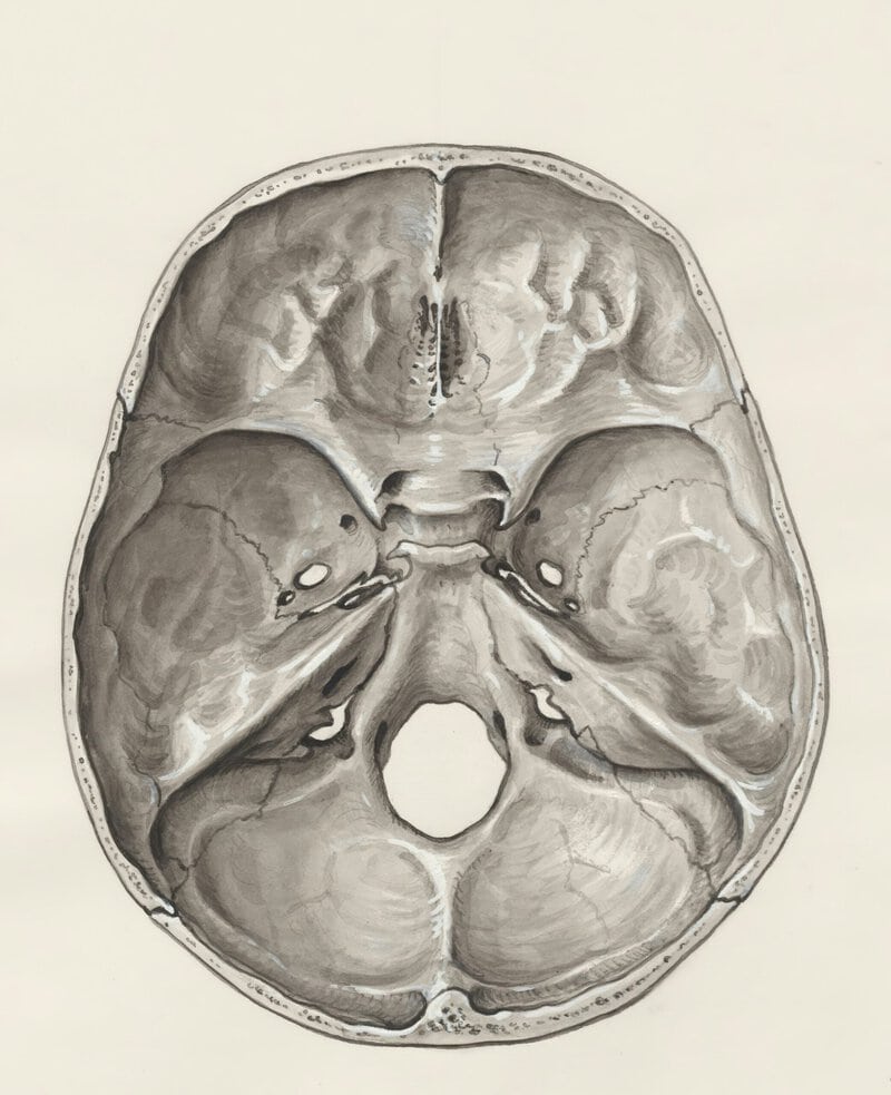
For most of medical history, the cerebrum collected nearly all the credit. It is the most visible part of the organ, the region most associated with intelligence, language, and consciousness, and the structure that expands most dramatically when comparing the human brain to those of other animals. The assumption, broadly speaking, was that bigger meant more important.
That assumption is now being tested. The human brain has three main regions — the cerebrum, the cerebellum, and the brainstem — and the relationship among them turns out to be far stranger and more elegant than a simple size ranking implies. Bulk does not equal control, and the brain’s division of labor may be one of biology’s most quietly remarkable arrangements.
What the Cerebrum Actually Is: Anatomy in Plain Language
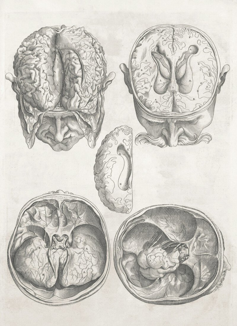
The cerebrum is the uppermost and largest part of the human brain. It is composed of two cerebral hemispheres — left and right — each of roughly equal size. Each hemisphere contains an inner core of white matter, the brain’s long-distance signaling cables, wrapped in the cerebral cortex: the deeply folded outer layer where most conscious processing occurs.
Those folds are not incidental. The ridges, called gyri, and the grooves between them, called sulci, dramatically expand the cortex’s surface area — estimated at roughly 2,500 square centimeters when unfolded — allowing far more neurons to be packed inside the skull than a smooth surface would permit. Evolution solved a packaging problem by crumpling the solution.
Popular culture frequently overstates left-brain versus right-brain differences. Research published in PLOS ONE in 2013 by Nielsen and colleagues at the University of Utah found no evidence that individuals preferentially use one hemisphere over the other for general cognitive function, although specific tasks such as language production do tend to be handled predominantly by one side. The two hemispheres are more cooperative than competitive.
A distinction worth keeping clear: the cortex is gray matter, dense with neuron cell bodies that process information; the inner white matter consists of myelinated axons — insulated fibers that transmit signals between regions rapidly. Both are essential, and neither functions without the other.
What the Cerebrum Does: From Movement to Memory
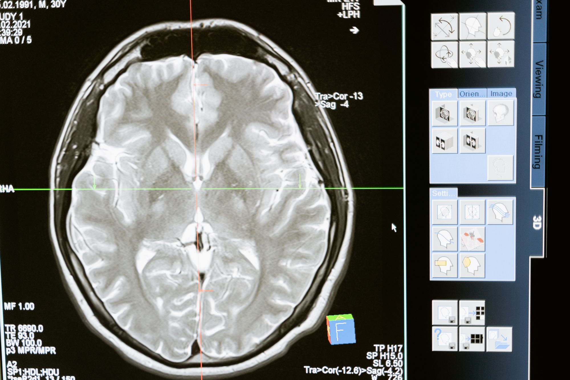
The cerebrum’s headline functions are extensive. It initiates and coordinates voluntary movement, processes all five senses, regulates aspects of body temperature through structures such as the hypothalamus, and underlies language, reasoning, and emotion — essentially everything commonly associated with distinctly human experience.
A concrete example makes this vivid. When a pianist reads a score and plays a chord, it is the cerebrum’s motor cortex that fires the movement commands, the somatosensory cortex that registers the pressure of fingertips on keys, and the auditory cortex that evaluates the resulting sound — a cascade completed in milliseconds, largely without conscious direction of each step.
Temperature regulation deserves a mention it rarely receives. The hypothalamus, a small but critical structure within the cerebrum’s lower regions, continuously monitors blood temperature and triggers responses such as sweating or shivering to maintain the body near its set point of 37°C. This function is easy to underappreciate until it fails, as it can in heatstroke or certain neurological injuries.
But the cerebrum does not operate alone. It relies on continuous signaling from the brainstem to sustain consciousness, and on the cerebellum to keep movements smooth and accurate. That dependency is the pivot point of this story.
Enter the Cerebellum: The Brain’s Compact Co-Pilot
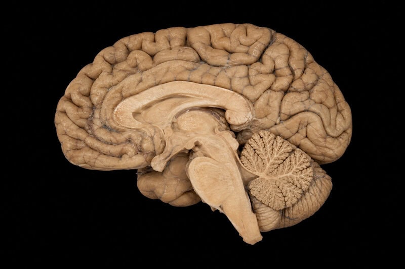
Here is the figure that stops most readers: the cerebellum makes up only about 10 percent of the brain’s total volume, yet it contains roughly 69 billion of the brain’s estimated 86 billion neurons, according to a landmark cell-counting study by Azevedo and colleagues published in the Journal of Comparative Neurology in 2009. The smallest major brain region is, neuron for neuron, the densest.
Its classical role is well established. The cerebellum — Latin for “little brain” — fine-tunes motor commands issued by the cerebrum, correcting errors in real time so that reaching for a coffee cup becomes a smooth arc rather than a jerky, overshooting lunge. This process, called motor coordination, becomes immediately obvious when the cerebellum is damaged: the result is ataxia, a poorly controlled, stumbling gait that resembles intoxication even in a completely sober person.
What remains an active and genuinely contested area of research is the extent to which the cerebellum also contributes to cognition and emotion. A 2019 review by Schmahmann and colleagues in the journal Neuron documented what they termed cerebellar cognitive affective syndrome in patients with cerebellar damage — deficits encompassing planning, language fluency, and emotional regulation — suggesting the cerebellum’s influence extends well beyond movement. Researchers caution, however, that clinical lesion cases establish correlation rather than causation. The evidence is compelling, but the precise role of the cerebellum in healthy human cognition remains harder to pin down.
Cerebrum vs. Cerebellum: A Functional Comparison
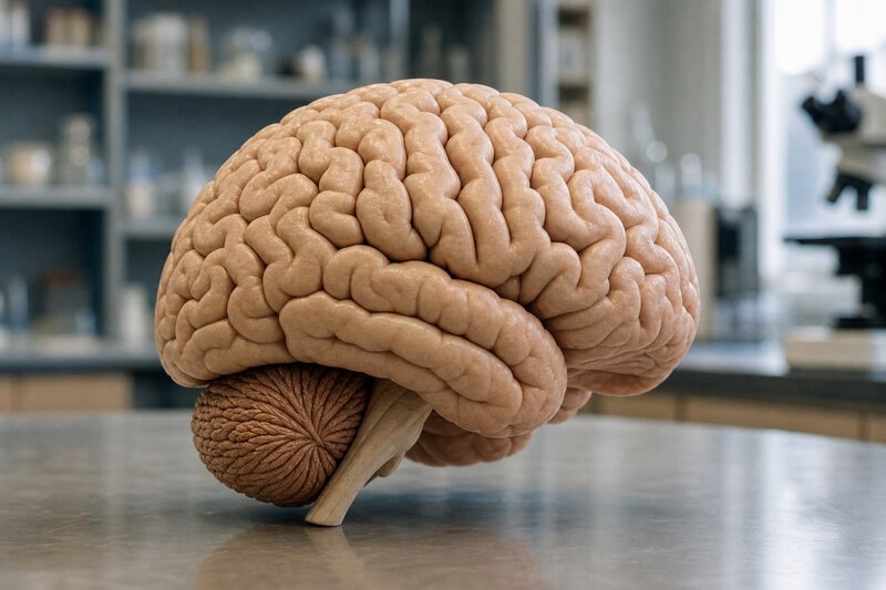
The cerebrum is the seat of conscious, deliberate cognition — language, decision-making, and the initiation of voluntary movement. The cerebellum operates largely below conscious awareness, acting as an automatic error-correction system that makes skilled performance feel effortless rather than labored.
An analogy illustrates the contrast clearly. A student driver consciously thinks through every turn, every mirror check, every gear change — a process heavily reliant on the cerebrum. An experienced driver executes those same maneuvers automatically and fluidly, barely aware of the individual steps. Neuroimaging research has shown reduced cortical activation as motor skills become automatized, a pattern consistent with the cerebellum absorbing a greater share of the workload as expertise develops.
The brainstem completes the three-part picture. As described in the NIH’s StatPearls reference database, the brainstem — the brain’s most evolutionarily ancient region — governs breathing, heart rate, and the sleep-wake cycle. It keeps the body alive so that the cerebrum and cerebellum can perform everything else. Without it, neither of the other two regions functions at all.
The point deserves a direct statement: the brain is not a hierarchy of importance measured by size. It is an interdependent system in which the cerebrum’s mass provides the computational canvas and the cerebellum’s neuron density may provide much of the precision brushwork.
What New Research Is Revealing — and What Remains Uncertain
A 2023 study published in Nature Neuroscience by researchers at the Allen Institute for Brain Science used single-cell RNA sequencing to catalog more than 3,000 distinct cell types across human brain regions. Among the findings the authors themselves described as unexpected was that the cerebellum contains a disproportionately large share of the brain’s overall neuronal diversity. That cellular variety may help explain how such a compact structure appears capable of contributing to such a wide range of functions.
One genuinely open question sits at the frontier of the field: whether the cerebellum contributes meaningfully to social cognition and autism spectrum conditions. Some researchers, including Jeremy Schmahmann at Harvard Medical School, argue that cerebellar circuits are involved in these domains. Others maintain that the evidence remains preliminary and that dysfunction in the cerebral cortex is the better-established explanation. Both positions are held by credible scientists working from real data; the question is not settled.
The stakes extend beyond academic debate. Understanding how these regions collaborate could reshape rehabilitation after stroke or cerebellar injury. If the cerebellum contributes to language recovery, therapies targeting only cortical regions may be leaving a meaningful tool unused. Getting the neuroscience right carries direct clinical consequences.
Epistemic honesty requires one further note: much of the evidence for the cerebellum’s expanded role in cognition comes from correlational research and lesion studies — observing what capacities are lost when a structure is damaged. Establishing direct causation in healthy human brains requires additional work using methods such as transcranial magnetic stimulation and high-resolution functional MRI. The picture is suggestive, not complete.
Why This Matters: Rethinking the Brain’s Chain of Command
The cerebrum’s status as the largest part of the human brain is not in dispute. Its roughly 85 percent share of total brain weight, its two hemispheres, and its billions of cortical neurons are among the most reproduced facts in neuroscience. But “largest” and “most important” have never been synonyms in this field, and the cerebellum’s extraordinary neuron density is a standing reminder of that distinction.
As National Geographic’s science coverage has noted, the human brain continues to yield surprises even after centuries of study. The emerging picture of the cerebellum is one of the more striking recent examples: a region long confined to a supporting role in movement now appears to be a broad-spectrum contributor to the very capacities that define human cognition.
The brain’s functional elegance may lie precisely in this arrangement — a large, flexible cerebrum handling conscious strategy, a compact, neuron-dense cerebellum handling automatic precision, and the brainstem sustaining life beneath them both. Each region enables the others; none is dispensable; none works in isolation.
The next time you catch a ball, thread a needle, or choose a careful word in a difficult conversation, the cerebrum started the process — but the cerebellum almost certainly helped it land. As connectome mapping and single-cell sequencing continue to advance, the boundaries between what each region “does” are likely to blur further, and the story of the brain’s largest part may prove inseparable from the story of its smallest.