When researchers injected a specially engineered virus into the brains of mice—designed to temporarily silence a small, specific cluster of neurons—the animals immediately lost their ability to filter out distractions. They became unusually reactive to irrelevant stimuli, struggled to sustain focus on meaningful cues, and displayed behavioral patterns strikingly similar to what clinicians observe in humans with ADHD. The culprit was a handful of ancient cells that most neuroscientists weren’t even looking at.
A Tiny Cluster With an Outsized Job
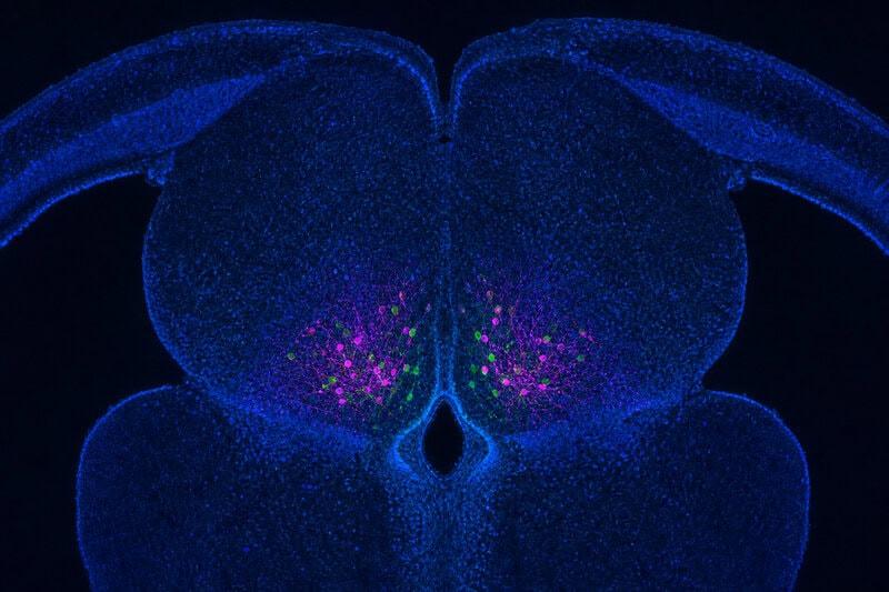
The neurons at the center of this discovery are called PLTi neurons—a small, discrete population tucked inside a subcortical brain region. “Subcortical” refers to structures that sit beneath the cerebral cortex, the wrinkled outer layer most associated with reasoning, language, and complex thought. These deeper structures are far older in evolutionary terms and are shared, in remarkably similar form, across a wide range of vertebrate species.
What makes PLTi neurons remarkable is not just their location, but their function. According to researchers at Johns Hopkins University, these neurons act as a built-in focus filter—not by amplifying the signals the brain wants to process, but by actively suppressing the competing noise it doesn’t. The distinction matters: PLTi neurons function less like a volume dial and more like a noise-canceling switch, selectively quieting irrelevant sensory information so that relevant signals can rise to the foreground.
The finding, reported in detail by Science Daily, represents a significant departure from where attention research has traditionally focused. For decades, the dominant scientific framework placed attention control primarily in the prefrontal cortex and parietal lobes—sophisticated, evolutionarily recent brain regions. The identification of PLTi neurons as a discrete attentional control point challenges that assumption at a fundamental level.
How Researchers Switched Off Focus in a Living Brain

To establish that PLTi neurons were causally responsible—not merely correlated with attentional behavior—researchers used a technique called chemogenetic inhibition. In plain terms, they engineered a virus to carry genetic instructions into the brain of a living mouse. Those instructions gave scientists an on-demand switch: when activated, PLTi neurons went quiet without requiring open surgery or causing permanent damage to surrounding tissue.
The behavioral results were immediate and measurable. Once PLTi activity was suppressed, mice began orienting toward irrelevant stimuli they would normally ignore. They struggled to sustain attention on cues that mattered. The distractibility was not subtle—it was a reproducible, consistent behavioral change observed across multiple animals and experimental conditions.
Control conditions confirmed this was not coincidence. Mice whose PLTi neurons remained intact maintained normal attentional filtering, demonstrating that the cells were causally driving focus, not simply accompanying it. That distinction is essential in neuroscience, where correlation is common and true causation is considerably harder to establish.
Mouse models carry particular scientific weight in this context because the brain region housing PLTi neurons is evolutionarily conserved—meaning its structure and core function are nearly identical across mice and humans. When a brain structure is preserved across species separated by vast stretches of evolutionary time, researchers have stronger grounds to expect that findings in animal models will carry relevance to human biology.
Why “Ancient” Matters: Evolution’s Oldest Attention System
In neuroscience, calling a brain structure “ancient” has a specific meaning: it refers to regions shared across species separated by hundreds of millions of years of evolution. When natural selection preserves a structure that long, it is generally because the function that structure performs is so fundamentally important to survival that organisms without it failed to reproduce. PLTi neurons, housed in one of these conserved subcortical regions, appear to serve exactly that kind of essential role.
This carries a striking implication: the basic architecture of attentional filtering was already in place long before complex cognition, language, or higher reasoning evolved. Attention, in this view, is not a luxury of sophisticated brains—it is a primitive survival tool, one of the earliest problems evolution had to solve. An animal that cannot distinguish relevant from irrelevant information in its environment cannot find food, avoid predators, or navigate safely. Selection pressure on that ability would have been intense and unrelenting.
The prevailing scientific consensus has long framed attention as primarily a cortical phenomenon, managed by the prefrontal cortex in concert with parietal and thalamic circuits. The PLTi neuron discovery does not dismantle that framework, but it adds a layer that was previously missing: a discrete, subcortical mechanism that may operate upstream of, or in parallel with, those cortical networks. Researchers are careful to note that identifying PLTi neurons as a key node does not mean attention is simple or entirely localized. The brain operates through interconnected networks, and this finding adds a critical piece without replacing the broader picture.
The ADHD Connection: A Parallel Worth Taking Seriously—and Carefully
The behavioral profile that emerged when PLTi neurons were silenced in mice—heightened distractibility, difficulty filtering irrelevant stimuli, impaired sustained attention—closely mirrors the defining features of ADHD, or Attention Deficit Hyperactivity Disorder. ADHD is a neurodevelopmental condition affecting an estimated 5 to 7 percent of children and adults worldwide, characterized by difficulty sustaining attention, impulsivity, and in many cases hyperactivity. It is currently understood to involve dysregulation of dopamine and norepinephrine signaling across prefrontal and subcortical brain circuits.
As noted by CHADD, the leading organization for ADHD research and advocacy, the PLTi neuron findings have drawn attention from clinicians and researchers interested in the disorder’s underlying neurobiology. The parallel between silenced PLTi neurons and ADHD-like behavior in mice is scientifically provocative—and appropriately cautious researchers are treating it as exactly that: a provocation worth investigating, not a conclusion worth announcing.
The study does not claim that ADHD is caused by PLTi neuron dysfunction, and no human PLTi neurons have been directly studied. The researchers have not proposed a causal link to any specific patient population. What they have done is identify a cellular mechanism that produces attention-related behavioral changes in an animal model—an early-stage mechanistic finding that, if replicated and extended, could eventually inform how scientists think about subcortical contributions to attention disorders. Translating that into human therapeutics would require years of additional research, independent replication, and ultimately rigorous clinical investigation.
What This Means for Neuroscience: A More Precise Map of Attention

One of the practical values of the PLTi neuron discovery is the precision it introduces. For years, attention research has grappled with diffuse network hypotheses—the idea that focus emerges from broad coordination across many brain regions, without a clear cellular mechanism to study or target. As Science Alert reports, the identification of PLTi neurons as a discrete attentional control point gives researchers something specific and concrete: a defined cell population whose activity can be measured, manipulated, and studied at cellular resolution.
The finding also fits into an evolving understanding of how attention actually works. Increasingly, neuroscientists conceptualize focus not as a single brain-wide state but as the outcome of two complementary processes: the suppression of irrelevant information and the amplification of relevant signals. PLTi neurons appear to be key players in the suppression side of that equation—a mechanism the field lacked a precise cellular handle on until now.
The methodological approach itself is significant. The combination of viral-vector delivery and chemogenetic tools allowed researchers to silence a tiny neuron population with high specificity and observe clean, interpretable behavioral outcomes. That level of cellular resolution is relatively new in systems neuroscience, and it opens the door to similarly precise investigations of other brain circuits whose functions have remained frustratingly opaque.
Important questions remain unanswered. How PLTi neurons communicate with the cortical attention networks already well-documented in human neuroimaging has not yet been established. Whether PLTi neurons are modulated by dopamine or norepinephrine—the neurotransmitters targeted by existing ADHD medications such as methylphenidate and amphetamine—is currently unknown. And whether equivalent neuron populations in the human brain behave in comparable ways will require neuroimaging studies and, eventually, post-mortem cellular analysis to answer definitively.
What We Know, What We Don’t, and Why It Matters

Scientists have identified a small cluster of ancient brain cells—PLTi neurons—that function as a focus filter in mice, and silencing them produces distractibility resembling ADHD, pointing to a previously underappreciated subcortical attention mechanism. That is the verified, peer-reviewed core of what has been found. It is a meaningful discovery. It is not a cure, a complete explanation of human attention, or a ready-made treatment target.
Independent replication across additional species, and eventually human neuroimaging studies capable of detecting analogous activity, will be essential before the scientific community can draw firmer conclusions about how directly this animal-model finding maps onto human attention and attention disorders. That process takes time, and it should—the history of neuroscience is littered with promising animal findings that did not survive the translation to human biology.
For general readers, the significance of findings like this lies in what they make possible downstream. Understanding the cellular machinery of attention at this level of precision is a prerequisite for developing better-targeted interventions—treatments that might act on specific neuron populations rather than producing the broad neurotransmitter changes that current medications use, which affect considerably more than attention alone. Every precise mechanism identified in neuroscience is a potential lever that future researchers and clinicians can pull.
The PLTi neuron finding does not solve attention. But it hands scientists a new lever—one rooted in some of the oldest circuitry the vertebrate brain has ever built, and one that may prove far more consequential than its tiny size suggests.