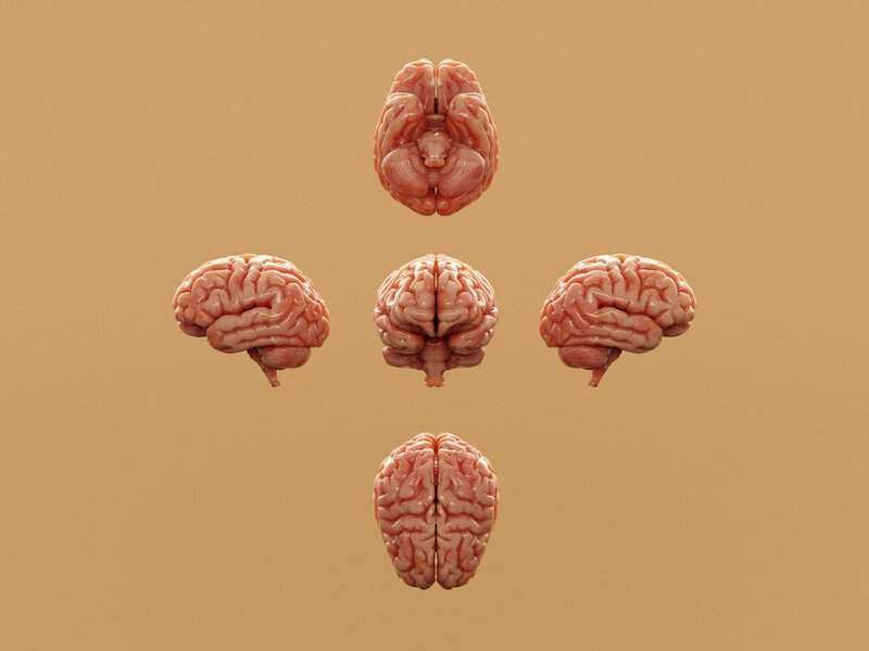
In advanced Alzheimer’s disease, the brain’s cortex can shrink so dramatically that its folded ridges — the gyri — reduce in size by up to 50%, the physical imprint of billions of lost neurons visible in autopsy after autopsy. Scientists have spent decades asking exactly what triggers that destruction, and for most of that time, the dominant assumption was surprisingly passive: neurons, the thinking went, simply suffocated under the accumulating weight of amyloid plaques and tau tangles. A wave of recent discoveries suggests the reality is far more deliberate — and far more complex.
Not Accidental Death: The Science of Programmed Cell Destruction
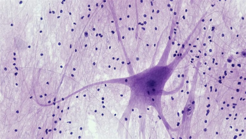
Biology has long recognized apoptosis — a tidy, controlled form of cell suicide the body uses to eliminate damaged or unwanted cells. But neurons in Alzheimer’s disease appear to die by a messier, more inflammatory route. A University College London study published in September 2023 identified that route as necroptosis: a form of programmed cell death in which a dying cell does not quietly dissolve but instead ruptures, releasing inflammatory signals that can damage neighboring cells. The UCL team demonstrated that this pathway was activated in a laboratory model of Alzheimer’s disease and directly led to the death of neurons — one of the first controlled demonstrations linking necroptosis to the disease.
That finding was then extended in an important direction. A study published in the journal Science took the question out of animal cells and into human ones: when human neurons were transplanted into mice carrying amyloid plaques, those neurons died specifically by necroptosis. That experiment established, for the first time in a human-cell context, a causal role for this pathway rather than a merely correlational one — a methodologically significant step that earlier laboratory models could not provide.
Necroptosis matters not just as a cause of death but as an amplifier. The inflammatory debris a ruptured neuron releases can activate the same fatal pathway in surrounding cells, raising the possibility that neuronal loss in Alzheimer’s spreads in part through a chain reaction of programmed self-destruction. That reframes the disease from a slow, passive accumulation of damage into something closer to a propagating signal — one that, in principle, could be interrupted at a defined molecular step.
A New Player: What Karyoptosis Adds to the Picture

Researchers part-funded by Alzheimer’s Research UK have identified a separate, previously underappreciated process called karyoptosis — in which a cell’s nucleus is expelled or degraded before the rest of the cell dies — that may play an important role in Alzheimer’s-related brain cell death. Where necroptosis describes how the whole cell ruptures, karyoptosis focuses specifically on the fate of the nucleus, the cell’s control center, and appears to occur earlier in the sequence of cellular collapse.
The distinction matters therapeutically. If scientists can interrupt the process by which a neuron loses control of its nucleus, they may be able to halt cell death before it becomes irreversible — a meaningful window of intervention that the older, passive model of Alzheimer’s neurodegeneration did not offer. The two processes are not necessarily competing explanations; researchers have raised the possibility that karyoptosis may precede and contribute to necroptotic rupture, making the sequence of destruction inside a dying neuron more ordered, and therefore more targetable, than previously assumed.
Both karyoptosis and necroptosis, however, remain emerging findings rather than established consensus. Researchers caution that confirming their roles in human Alzheimer’s brains — not just laboratory models — will require larger, longitudinal studies before either pathway can be considered a validated therapeutic target.
Toxic RNA: A Third Mechanism Identified at Northwestern

A third line of evidence arrived in January 2024, when researchers at Northwestern University announced the identification of short strands of toxic RNA molecules that contribute to brain cell death and DNA damage in Alzheimer’s disease. RNA, or ribonucleic acid, normally ferries genetic instructions from DNA to the cell’s protein-making machinery. The toxic variants identified by the Northwestern team appear to interfere with that process, causing cellular stress and triggering DNA damage in neurons — a mechanism distinct from both necroptosis and karyoptosis, and one that the team described as the first discovery of its kind in this disease context.
The finding adds a third layer to the emerging picture of how Alzheimer’s kills neurons. Amyloid plaques and tau tangles may set the stage, but toxic RNAs and programmed death pathways like necroptosis and karyoptosis now appear to be among the actual executioners. The Northwestern study was conducted in laboratory models, and the researchers noted that translating these findings into potential therapies will require validation in human tissue and eventual clinical trials — a timeline that remains uncertain but that the discovery now makes worth pursuing.
The Brain’s Failing Cleanup Crew
None of these cell-death pathways operates in isolation. According to the National Institute on Aging, a type of glial cell called microglia — the brain’s resident immune and waste-disposal system — normally engulfs and destroys harmful debris, including the misfolded proteins implicated in Alzheimer’s. When microglia fail to clear amyloid plaques and other toxic waste efficiently, the accumulating debris creates conditions that appear to prime neurons for the very death pathways researchers are now documenting.
Some scientists hypothesize that the newly described cell-death mechanisms and microglial dysfunction are not sequential events but a feedback loop: dying neurons release inflammatory signals that impair microglia, which then fail to clean up the debris from those dying neurons, accelerating further neuronal death. This interaction between neurons and their supporting glial cells is an active area of investigation, and researchers caution that the causal direction of the relationship — whether microglial failure precedes neuronal death or follows it — has not been definitively established in human Alzheimer’s disease. Resolving that question will be critical to determining at which point in the cycle a drug could most effectively intervene.
What This Means for Treatment
Identifying necroptosis, karyoptosis, and toxic RNA as candidate mechanisms in Alzheimer’s brain cell death shifts the therapeutic conversation in a concrete way. Rather than focusing exclusively on clearing amyloid plaques after they have already formed — the strategy that has defined most Alzheimer’s drug development for two decades — researchers can now explore compounds that block specific steps in these death pathways upstream of neuronal loss, intervening before damage becomes permanent.
The practical implications break down into several distinct avenues:
- Necroptosis inhibitors — molecules that block the proteins driving that cell-death cascade — already exist in experimental form for other diseases, giving Alzheimer’s researchers a potential shortcut rather than starting drug development from scratch. The key question is whether those compounds can cross the blood-brain barrier at therapeutic concentrations.
- Karyoptosis-targeted therapies could aim to preserve nuclear integrity in neurons showing early signs of stress, potentially halting the chain of events before a cell ruptures and triggers inflammation in neighboring tissue. Because karyoptosis may precede necroptosis, this window could offer an earlier intervention point.
- RNA-silencing approaches that neutralize harmful RNA strands represent a separate avenue. This class of therapy has already achieved clinical success in other diseases — silencing mutations that cause spinal muscular atrophy, for instance — and may be adaptable for Alzheimer’s if the specific toxic RNA targets identified at Northwestern can be validated in human brain tissue.
Experts emphasize that all of these approaches remain at early, preclinical stages. None has yet been tested for safety or efficacy in human Alzheimer’s patients. The history of Alzheimer’s drug development — marked by many compounds that succeeded in mice but failed in human trials — warrants measured expectations about timelines, even as the underlying science becomes more mechanistically precise.
A Convergence That Changes the Question
Taken together, the UCL necroptosis findings, the Alzheimer’s Research UK-funded karyoptosis work, and the Northwestern toxic RNA discovery represent a meaningful shift in the field. Research into neuronal cell-death mechanisms in Alzheimer’s disease has historically been fragmented, with different laboratories pursuing different hypotheses in parallel and rarely converging on shared conclusions. What is notable about this recent cluster of findings is precisely that convergence: each, from a different experimental direction, points toward neurons as active participants in their own destruction rather than passive victims of external damage.
The cortical shrinkage visible in Alzheimer’s autopsies — up to 50% reduction in gyral size in some regions — now has a set of candidate mechanisms to explain it. Those mechanisms are active, programmable, and in principle interruptible. Confirming them in large-scale human tissue studies, and determining whether blocking them early enough in disease progression can meaningfully preserve cognition, is the work that now lies directly ahead.
The question for Alzheimer’s science has shifted. It is no longer simply “what kills neurons?” It is “can we intervene at the exact moment a neuron commits to dying?” — and for the first time, researchers believe they are close enough to that moment to design experiments that could answer it.